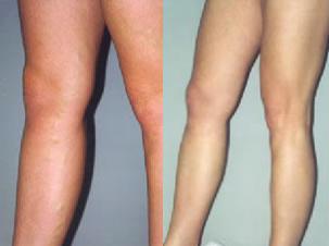
I am 35 and I have new, smooth, shapely legs. “Why new?”, one may ask. Because I can show them just as I used to do it when I was younger.
When I was a young and beautiful girl, I used to wear mini skirts. They were fashionable, at that time, and I was not ashamed to show my beautiful, shapely and smooth legs. Even during winter I was not scared of the temperatures below zero or not to mention snow, and I simply wore my beloved mini skirts. My teachers used to nag all the time about the length of my and my friends’ skirts, or rather their shortness. However, we knew how to deal with it. The skirt was usually longer, up to the knees, but after having left home or school, we pulled it up so that it would scarcely cover the buttocks. Unfortunately, with age my legs were not as beautiful as they used to be and, consequently, I did not show them so eagerly. There started to appear gradually little widened blood vessels on the skin in the region of thighs and calves that resembled little webs. Little venules, almost invisible at the beginning, were becoming wider and wider and did not look nice. I gave up wearing short skirts, and started wearing long ones and trousers. I was a little worried about the condition of my legs.
I remembered very well that my grandmother suffered from varices of the lower legs. She did not undergo any operation as she was scared of having any very much. Even though she was not very old, she used to sit on the coach very often with swollen and painful legs, especially in the region of ankles, and she asked me to massage them a little. While massaging them, I could feel under thick tights widened and large varicose veins.

My professional job requires sitting long hours in front of a computer screen. So, many times I could feel a kind of heaviness in my legs, and in the evening I noticed slightly swollen ankles. I read a little about varices in the books available. I knew it was a hereditary disease and I could expect it somewhere in the future. I even tried to change my lifestyle a bit. At work I got up and do some physical exercises from time to time. I did some climbing up on my heels and toes, short walks in the office and, during the breaks, I put my legs higher. I tried not to wear socks up to my knees or socks, generally, that would squeeze my calves. I simply wore tights or naked legs. In summer on hot days, I did not wear high-heeled shoes or shoes that would constrain or squeeze my feet, but rather flip-flops and loose comfortable shoes. I did not go to solarium, sauna or take hot baths.
My tragedy, however, started when I got pregnant. At the very beginning everything was as it used to be. Nevertheless, when my belly grew bigger and bigger, so were the changes in my legs. Little varicose veins changed into big ones. Moreover, new ones also appeared, earlier not present at all, which with elapse of time changed into bigger widened blood veins. I even scheduled an appointment at the doctor’s. First, my gynaecologist examined me and recommended a surgical consultation. The surgeon told me that in such cases, with some genetic endowment, sitting or standing character of my job, during pregnancy, varices either exacerbated or started to appear. Unfortunately, he could not help me due to the state I was in. He recommended me a few additional exercises, comfortable shoes and patience, and another visit, this time after the delivery. I gave birth and raised my daughter. Unfortunately, the problem of my legs came back. Still, I was not an old woman, and I would eagerly uncover my legs during summer, on the beach or at the swimming pool.
So, again I visited the surgeon. He prescribed me having ultrasonography examination, the so called Doppler’s test to measure the capability of the venous system of the lower legs. It was the examination from which he conditioned the size of the surgery because, as he warned me, the operation was unavoidable. My varices were not any longer a cosmetic problem, but after all a healthy one as varices might result in very serious complications such as for instance, pulmonary embolism which, finally, could end up with death in the future. Scary, isn’t it? I did not have any choice. I underwent the prescribed medical examination and having all the results in my hand I arrived in the right place and in the right time. The surgery was performed under a general anesthetic. The surgeon informed me that the surgery consisted in removing incompetent superficial veins, one main saphenous vein and other smaller lateral veins changed due to varices. When I woke up the next day, my both legs were covered with elastic bandages. In the region of the groin, there were little dressings covering slight cuts each being 3cm long. After the morning doctor’s visit and change of the dressing, I went back home. During the following two weeks, my legs changed colors into violet, navy blue, green and yellow. In the places where I could see bruises, the body was hard and painful. However, after two weeks everything came back to the norm. There were just few places where I could feel the pain while pressing the place harder. There were, however, no visible and protruding “grape bunches” as before. Few more weeks later I totally forgot about my varices. Then, beautiful and warm spring came, and I started showing my legs and, again, I convinced myself to wear mini skirts.
Judyta
Surgical treatment of varices – radicalism and cosmetics
There are 1,8% of women and 1,1% of men aged between 30-39 who suffer from varices. The frequency of the disease increases with age and refers to 20,7% of women and 24,2% of men over the age of 70. In Poland, it usually attacks women, namely 69% of their population suffer from varices.
Varices cause either objective problems like swelling, discolorations, echemas and ulceration or is connected with more serious complications like inflammation of superficial veins, deep vein thrombosis or pulmonary embolism.
It also causes serious cosmetic defects.
The treatment of veins changes can be either preventive or surgical ones. However, in the case of fully developed disease, only surgical treatment allows to obtain the desired therapeutic and cosmetic effects.
The venous system of the lower legs consists of superficial veins and deep ones connected with each other by means of perforating veins and the outlet of saphenous and small saphenous vein. Deep veins of the lower legs run under the fascia, and in doubled number accompany the arteries. Superficial veins run under hypodermis tissue. They are accompanied by sensory nerves fibers and lymph vessels. In the case of a healthy man, 90% of blood flows from the lower limbs to the heart by means of deep veins, and only 10% through the superficial system.
The moment muscles contract during any movement of the leg, blood pressure increases inside the fascial sheaths up to 200 mm Hg. Then, it squeezes blood out of the little veins in the region of the muscle, and also from bigger veins which run through fasciae. Blood goes through venous valves that give it a proper direction. During muscle diastole, blood pressure decreases in the veins, however, properly working valves unable blood regurgitation. This way deep veins have temporarily low blood pressure that makes them be able to suck blood from little muscular and superficial veins. A normal flow of blood from superficial veins to the deep ones is assured by perforating veins and the outlets of saphenous and small saphenous veins together with their valves.
Varices of the lower limbs.
Varices are morbidly changed veins with their permanent baggy and fusiform widening and prolonging with a fixed incompetence of the valves of the superficial and perforating veins. These are women who suffer most often. In 85% of the cases they refer to the system of small saphenous vein. The factors responsible for varices most often are as follows:
- heredity
- race (most often occur in the case of white race)
- overweight
- undergone pregnancies
- hormonal treatment
- type of activity (sitting or standing job)
Currently, there are two theories presented most often concerning the reasons of varices, namely hemodynamic and the theory of incompetence of a vein wall.
- Hemodynamic theory; the main factor contributing to creating varices is hydrostatic blood pressure in veins. The pressure is to result in valves damage and the following widening of veins. By the same token, blood regurgitation takes place.
- The theory of incompetence of a vein wall is based on some genetic premises.
The creation of varices is explained in terms of degeneration of collagenous fibers and the atrophy of muscular cells of the internal and external membrane. Varices widening result in valves damage which, consequently, results in reverse of blood.
In physiological conditions, blood always flows from superficial saphenous veins to the deep ones in two ways. One, through the outlet of the saphenous or small saphenous or perforating veins. This blood flow is supported by a muscular pump (the moment a muscle diastoles, blood is sucked in from the superficial veins to the deep ones).
In the case of damaged valves, blood is squeezed under a huge blood pressure from deep veins into the superficial ones. The created reflux causes high blood pressure in the superficial veins which may increase even to 200 cm H2O. It results in the development of widened superficial veins, i.e. varices.
Depending on the degree of venous system incompetence, one may differentiate the following stages of the disease.
Zero degree Lack of clinical ailments. The sick wear the burden of genetic premises passed down by their family. The stem of saphenous vein is slightly visible through the skin.
First degree Light symptoms. The sick declare the feeling of legs heaviness, temporal swelling in the region of the ankles, growing in the afternoon. The stem of saphenous vein can be widened.
Second degree Medium serious stage. Past history as in the first degree. Widening of the saphenous vein on a longer distance. Massive hypodermic varices, discoloration in the region of the ankles. The symptoms of reverse leak in the outlet of saphenous and small saphenous veins confirmed by Doppler’s examination.
Third degree Serious stage. Skin complications connected with the damage of valves, massive varices and swellings.
The doctor deciding on the surgery should know since when the symptoms have been occurring, whether their occurrence was connected with undergone pregnancy, whether the patient suffers from swellings and crural cramps, whether suffered in the past from the symptoms typical of inflammation of veins. It is also important whether the patient takes hormonal medications (contraceptives).
While examining a patient, the doctor examines carefully the way varices run, estimates whether there are any swellings, skin discoloration or ulceration, and makes many trial activities.
The performance of duplex Doppler’s test with colorful marking of the flow is extremely important. It helps to estimate the condition of the deep veins as well as it shows the places where the reverse of blood takes place. Thanks to this examination the surgical treatment is more precise and effective.
The treatment of varices
The aim of the treatment is to eliminate all the places of the leak of blood from the deep system to the superficial one as well as the removal of the veins changed by the disease. The treatment can be either preventive or surgical one. In the case of developed varices, only the surgical treatment allows to obtain proper therapeutic and cosmetic effects.
One of the stages of the operation is the removal of incompetent and widened saphenous vein. Consequently, a slight cut is made in the groin and in the region of medial ankle. The application of a proper technique results in almost invisible scars.
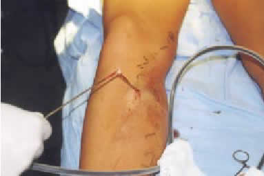
The further stage of the operation consists in the removal of varices and their branches. One of the most attractive ways to do so is the Müler’s method which consists in making 2-3 mm long incisions and removal of a single varix by means of a special hook. Such incisions can be done in a number of places along a particular varix removing it on the whole length. If the cuts are made in accordance with skin lines and closed with Steri-Strip plaster, the scars after 6 months are almost invisible. An improved method of this operation is the operation with a cryosurgical probe.
At the very end of the probe, a very low temperature is sustained up to ’80C degrees. The area of freezing around the probe equals 3-6 mm. The moment the probe freezes of, it sticks to the varix enabling its removal. This way, from one slight incision, a large number of varices can be removed in all directions on the length of 20 cm. The cut is usually ended with a Steri-Strip plaster. It allows to obtain a very good cosmetic effect.

The cryosurgical probe allows not only to remove varices (cryostripping). Thanks to it, the closure of vein vessels is possible to make, the so called cryobliteration. This method is usually used in the case of people with high operational risk, at whom the removal of varices could be dangerous to their health or lives.
Cryosurgical operations of varices have the following advantages:
- The possibility of a more precise removal of varices without the surrounding tissues.
- The possibility of operation after having undergone sclerotherapy
- The possibility of removal an incompetent saphenous and small saphenous veins without any additional skin cuts.
- The possibility of operation with the presence of little trophic changes
- Beneficial aesthetic effects
- Smaller number of the required skin cuts
- Shorter time of operation
The appliance of the operational technique is connected with the necessity of having very specialized equipment as well as the surgeon is required to have proper job experience at operations with the use of cryosurgical probes.
However, the advantages of this method, especially very good cosmetic effects together with its radicalism make that it is a strongly recommended method of the treatment of varices.
Jerzy Kolasiński, M.D. Ph.D.
The method of cryosurgical operation of varices has been used in Hair Clinic Poznań for many years. The work devoted to its use written by the group of surgeons from Hair Clinic Poznań and was published in THE AMERICAN JOURNAL OF COSMETIC SURGERY.
The author of the article led a course devoted to this method during 16th Meeting of the American Academy of Cosmetic Surgery w Orlando, USA in January 2000.
Sclerosis application in the case of incompetence of the lower legs veins
Sclerotherapy is one of the therapeutic methods used in the treatment of veins chronic incompetence. This well known, for many years, method in Western Europe supports the treatment of the lower limbs varices.
Sclerotherapy consists in purposefully induced inflammatory reaction of a blood vein wall changed due to varices which, finally, results in. What is obtained is the closure of the vessel lumen. The injected medication irritates the inner membrane bringing about vasoconstriction. An additional mechanism, which closes the vessel of 2/3 of its transection, works on a nervous way. Thanks to such an irritated and narrowed vessel, a thrombus is created, which next undergoes fibrosis and scaring. The vessel closes and stops being patent.
Varices of the lower legs, ulceration over incompetent vessel plexuses as well as incompetence of the perforating veins in embolism syndrome. The postphlebitis syndrome is a indicator to perform sclerosis.
The most popular medications used to close vein vessels are as follows: Scleremo, Variglobin, Varicocid, Aethoksysclerol. Unfortunately, not each of those medications is available in Poland.
The technique of medical treatment in the case of varices obliteration and venulactasies of the lower legs consist in injecting obliteration medication directly to the visible just under the skin changed vessel.
Recently, however, preparations in the form of foam instead of water solution have been used more eagerly. The main advantage of a foam is a slower inactivation of the obliteration medication before it reaches the aim as well as the transport of the active substance in the direction of vessel wall is more effective. The application of foam allows to apply a lower level and smaller amount of the medication, which, consequently, increases the safety of its use. Its efficacy is much longer as longer is the time of its working in a particular region.
In order to perform sclerosis, it should be remembered about numerous anatomical types of the venous system. Before performing sclerosis of a greater vein axes, ultrasonography examination should be performed as well as Doppler’s test of the lower legs or, even better, the examination with a colorful picturing flow, the so called color Doppler’s test.
The aim of the treatment is the closure of varices, lowering vein pressure after a physical effort and prolonging the time of blood inflow to the veins after muscular effort was made.
The sclerosis of incompetent superficial veins is least efficient in the case of coexisting reflux, i.e. blood flowing backwards into the deep system as most often it may result in redrainage of the vessels. The conclusion is as follows. In the case of patients suffering from blood reflux to the deep veins, more effective forms of treatment, namely surgical one, should be applied, not sclerosis. Additionally, such patients should wear tights or up to the knees socks of gradual compression during the convalescence after the surgery.
In the case of obliteration of huge vein axes, specially adjusted pressure sponges are used as well as compressive tights are recommended, and in the case of obliteration of small vessels, an elastic bandage squeezing the places of medicine administration place is enough. Such a dressing is worn from 24 up to 72 hours. In this period of time the creation of thrombus takes place that usually lasts till 18th day after the surgery. Only then, the final result of the obliteration therapy is visible. After the surgery, the sick should avoid having hot baths as well taking medications that lower blood coagulation.
The most often and difficult treated cases are slight and little widened skin vessels, the so called vessel webs; blue ones contain venous blood (venulactasies) and the red ones contain arterial blood (teleangiectasies). Blue vessels are easier to close, but they also may be the problem, especially, when they have a number of vertical and horizontal connections with deep vessel plexus or even huge deep veins or muscular ones.
The surgery of sclerosis should be performed several times, with two days breaks, especially, at more expanded changes. At 30% of the cases very good results are obtained (the disappearance of widened vessels) and good results at 40% (almost total disappearance of the widened vessels). Complications occur extremely rarely (generally about 0.5%), and most often are skin necrosis and trophic discolorations.
Małgorzata Kolenda, M.D. Ph.D.